Students Present at SPIE in Houston, TX

On February 11 and 12, Kristina Landino (BME) and Shuyue Guan (BME), advised by Dr. Murray Loew (BME), each presented a paper at the SPIE Medical Imaging conference, held in Houston, TX

K. Landino and M. Loew. “Comparing salience detection algorithms in mammograms.” (presented by Kristina Landino).

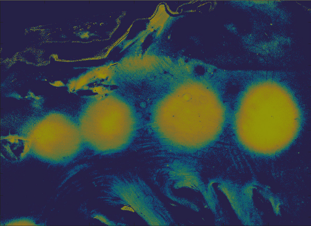
S. Guan, H. Asfour, M. Loew, N. Sarvazyan, and N. Muselimyan. “Lesion detection for cardiac ablation from auto-fluorescence hyperspectral images.” (presented by Shuyue Guan)

*Shuyue Guan presenting his poster at SPIE 2018

*Kristina Landino presenting her project. Photo credit to Dr. Ken Hanson

Student presents at 46th Annual IEEE AIPR 2017 Workshop

Our PhD student, Shuyue (Frank) Guan, has attended the 46th Annual IEEE AIPR 2017: Big Data, Analytics, and Beyond in Washington DC. Frank gave a presentation about breast cancer detection using transfer learning in the convolutional neural networks.

The Applied Imagery Pattern Recognition (AIPR) workshop sponsored by IEEE is to bring together researchers from government, industry, and academia across a broad range of disciplines. The Big Data Analytic domains represented at AIPR 2017 include computer vision, remote sensing imagery, medical imaging, and robotics and tracking, with a focus on machine learning and deep learning.

Here is a brief summary of Frank’s project and presentation:

Traditional mammographic detection based on the computer-aided diagnosis (CAD) tools rely on manually extracted features, but hand-crafted features have a variety of drawbacks such as domain specific, and the process of feature design can be tedious, difficult, and non-generalizable. An alternative method for feature extraction is to learn features from whole images directly through the Convolutional Neural Network (CNN), however, training the CNN from scratch needs a huge number of labeled images. Such a requirement is infeasible for mammographic tumor images because they are difficult to obtain, diseases are scarce in the datasets, and expert labeling is expensive. A promising solution is to use a limited number of labeled medical images to fine-tune a pre-trained CNN model, which has been trained by very large image datasets from other fields. This approach is also called transfer learning. In fact, some results of transfer learning are counter-intuitive: previous studies show that the features learned from natural images could be transferred to medical images, even if the target images greatly differ from the pre-trained source images.

Using mammographic images from the two databases, we tested 3 training methods: (1) trained a CNN from scratch, (2) applied the complete VGG-16 model to extract features from input images and used these features to train a classifier, (3) updated the weights in several last layers of VGG-16 by back-propagation (fine-tuning) to detect abnormal regions. By comparison, we found that the method (2) is ideal for study. Then, we used method (2) to classify regions: benign vs. normal, malignant vs. normal and abnormal vs. normal from DDSM. Our results show an average accuracy of about 90.5% for abnormal vs. normal classifications on mammography and the AUC is 0.96 are competitive. Our best model could reach 95% accuracy for abnormal vs. normal case. Compared with recent studies, we used much more images for training, different pre-trained model and simpler classifier.

This study shows that applying transfer learning in CNN can detect female breast cancer from mammographic images. And, training classifier by extracted features is a fast way to train a good classifier in transfer learning.

Students Attend BMES Annual Conference in Phoenix

Our students Aidan Murray, Shannon Toole, Zainab Mahmood and Nada Kamona have participated and presented their projects in the Biomedical Engineering Society Annual Conference held on October 14th in Phoenix Arizona. Aidan and Shannon presented on cluster and quadrant analysis for thermographic breast cancer detection. Zainab presented her work on developing an automated segmentation algorithm for thermal breast images. Nada presented her summer project at the FDA on the variability of image texture quantification in simulated medical imaging systems. Check their projects here and the photo gallery here.

The annual meeting is held annually by The Biomedical Engineering Society (BMES) and is the home for more than 2000 scientific presentations, platform sessions, exhibit hall and career fair, offering networking and career development opportunities for students and professionals.

Congrats to our students on their accomplishments!

Our Students Discuss their Internship Experience in Summer 2017

This past summer, many of our students had the opportunity to get their foot in the door of prestigious organizations, such as Singh Center for Nanotechnology, U.S Food and Drug Administration (FDA), and GW’s Nanotechnology Fellows Program. Here is what Caitlin, Nada and Zainab share about their accomplishments:

Caitlin Carfano:

This summer I learned more than just information about the project I worked on, but also how to operate advanced equipment such as an Atomic Force Microscope. I regularly conducted research in the Singh Center for Nanotechnology surrounded by state of the art equipment and working with graduate students in my lab. I really enjoyed being able to work so closely with my mentor, Annemarie Exarhos, because I got to ask ample questions about research and I received numerous tips on creating and executing presentations. I also participated in a poster session symposium with other REU students and Penn scholars!
After learning about quantum technologies and how rapidly this field of study is advancing, I want to continue working in this field. Participating in this program also opened my eyes to all the other scientists and engineers doing quantum engineering research (I had a stimulating conversation with my eye doctor about quantum optics patents that he is working on!). I have always wanted to help others by pursuing a health/medical related career. After discovering this growing field and its incredible potential, I would be interested in research that applies quantum technologies to medical imaging devices.

Nada Kamona:

This summer, I was an ORISE Research Fellow at the U.S Food and Drug Administration (FDA) in the Division of Imaging, Diagnostics, and Software Reliability (DIDSR). Image analysis techniques, such as computer-aided diagnosis or radiomics, often rely on quantitative measurements of textures as input to characterize disease status. Ideally, texture features should discriminate different textures, and should be robust across a range of patient characteristics and image acquisition conditions. This study aims to identify such texture features through simulation. In my project, I worked on the variability of image texture quantification in simulated medical imaging systems, by examining the repeatability and reproducibility of 35 texture features to noise and spatial resolution using a library of textures that we generated. Not only has my positions allowed me to learn about quantitative imaging on a more in-depth and practical level but it has also taught me valuable critical thinking and problem solving skills. Working together with my supervisors at the FDA has helped me become a well-rounded scientist who is capable of both working by another’s direction and being an independent thinker.

Zainab Mahmood:

This past summer I conducted research under the Nanotechnology Fellows Program, a program designed to introduce students to nanotechnology, cutting-edge research, and GW’s new nano facilities. Through this program students attended lectures, seminars, and received hands-on cleanroom training. They received training on how to operate tools needed in nanofabrication and characterization processes such as the electron-beam lithography (EBL) tool, SEM, AFM, confocal microscope, probe station, and thermal evaporator. For my research project, I fabricated micro-scale gold contact design on graphene ribbons using Raith CAD software, electron beam lithography, oxygen plasma etching, and physical vapor deposition using a thermal evaporator and characterized electrical properties of graphene using four-point probe analysis and Raman spectroscopy.