
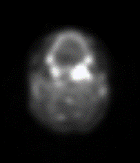
Quantitative dynamic thermal imaging as an adjunctive biomarker in breast cancer detection
This is a translational project to develop and refine a clinical protocol for thermal (infrared) imaging of the breast, to develop algorithms for automated analysis and classification of the images, and to use pilot studies to assess the clinical feasibility of dynamic (time-based) thermal imaging as an adjunct to mammography for detection of breast cancer. We will design algorithms for making inter- and intra-breast comparisons; they will incorporate our thermal and deformation models to establish criteria for defining regions of suspected abnormality. Methods for evaluation of changes in temperature with time, across the surfaces of the breasts, are expected to lead to improved detection of deep masses. We will continue to recruit volunteer subjects, all scheduled for breast surgery, from the Breast Care Center population, image them, make inferences using our software, and compare to “truth” as provided by the Center. We will report results as sensitivities and specificities (as related to sizes and depths of the lesions) and as receiver operating characteristic (ROC) curves.
Collaborators: GW Medical Faculty Associates, Breast Care Center
New generation of catheters for treatment of atrial fibrillation
Atrial fibrillation (AF) remains the most commonly occurring cardiac arrhythmia. It is associated with a lower quality of life and a higher rate of morbidity and mortality. One of the main options to treat atrial fibrillation is to ablate abnormal sources of electrical activity using percutaneous radiofrequency (RF) catheters. RF lesions isolate the spread of electrical activity from the sites of abnormal activity, including sleeves of the pulmonary veins. To date, there are limited means for real-time monitoring of tissue injury during RF ablation procedures. This project will create a new generation of imaging catheters based on spectral changes in tissue autofluorescence caused by thermal damage. We will acquire emission from multiple bands within the 380-600nm autofluorescence range and use post-acquisition principal component and other kinds of analyses to reveal thermal damage to layers of endocardial collagen and underlying atrial muscle. The UVA multispectral catheter will be then used to examine and visualize RF ablation lesions made via percutaneous access in live large animals such as pigs. We will then test it in freshly excised donated human atrial tissue. The catheter will then undergo extensive documentation, verification, and validation in order to be approved for use in a human study. Our ultimate long term goal is to develop a new generation of ablation catheters enabling real-time visualization of ablated cardiac tissue. This technology will pave the way for easier, faster, safer, more cost-effective, more reliable and minimally invasive AF ablation procedures
Collaborators: GW School of Medicine and Health Sciences, Nocturnal Product Development, LLC
Funding: National Institutes of Health
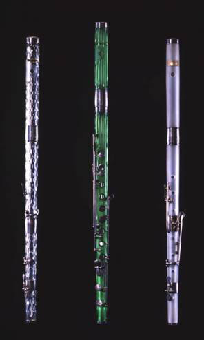
Glass at Risk: simple tools for detecting unstable glass in 19th century cultural heritage collections
This project supports interdisciplinary, collaborative research into the development of an “identification tree” that uses ultraviolet (UV) light tools as a simple, first level examination step, and leads to methodical application of other non-invasive techniques that allows identification of unstable and deteriorating types of glass in cultural heritage collections. The research will use several groups of objects to develop separate databases that establish UV response patterns and their correlation to elemental composition: (1) glass flute musical instruments made by Claude Laurent and studio in Paris in the first half of the 19th century; (2) cover glasses on encased 19th C photographs; and (3) glass substrates used for 19th century photographs. This work will go hand-in-hand with research that refines our understanding of typical 19th century glass types and their respective stability, which, in the case of common potash glass, is extremely poor. The identification tree will therefore aid caretakers of cultural heritage first and foremost by providing simple sorting mechanisms that identify at-risk cultural heritage, even when deterioration is not yet evident. This examination protocol and methodology, which may be different for separate types of glass-containing artifacts, will be simple, low-cost, and tailored to end-users who are not scientists at the first level, but will expands to include progressively more sophisticated techniques for detailed study and higher degree of confidence by the museum professional.
Collaborators: Library of Congress, Catholic University of America
Funding: National Endowment for the Humanities
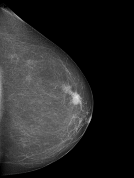
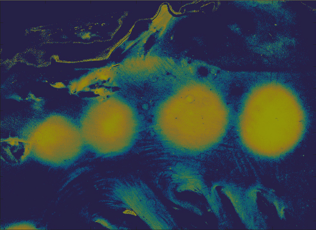
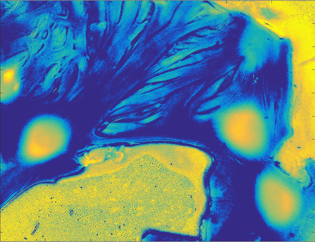
Comparing salience detection algorithms applied to mammograms
Salience in imaging is defined as the extent to which an object in an image catches the eye of the viewer. Currently, several software packages exist that calculate salience using a wide range of models and implementations. For example, in several of the models examined here, the software creates a series of maps for individual salience features like orientation and intensity, and then combines those individual feature maps into an overall map of salience for the entire picture.
Differences exist between these models in the way feature maps are calculated, in the way they are integrated into a single map, and in the ways that various types of noise are minimized. Differences also exist in the use of mid-level and high-level features like horizon line detection and facial and person recognition software. In other models, neural networks were used to create a series of layers, each of which transformed the data and found the most salient points in an image. This work compares 15 models, including our own algorithm, on the basis of speed and mass-detection accuracy, when applied to a common database of mammograms.
Development of a novel radiomics platform to predict outcomes in advanced head and neck cancer
Quantitative medical imaging (radiomics) offers the opportunity to characterize head and neck squamous cell carcinoma (HNSCC) in ways that may allow prediction of patient outcomes after radiation therapy. We intend to develop computational tools that will use data from PET scans to predict recurrence of tumor in patients treated with radiation therapy. Current progress involves determining whether information present in the heterogeneity of tumor regions in pre-treatment PET scans of Head-and-Neck Squamous Cell Carcinoma (HNSCC) patients and in their gene mutation status can measure efficacy of radiation therapy in their treatment. We’re investigating the efficiency of texture measures in quantifying the tumor heterogeneity, which can be used to predict recurrence status. Combined with patients’ gene expression patterns, texture measures can substantially improve therapy response prediction scores. This can help reduce the number of patients who are unnecessarily exposed to radiation. Our radiogenomic workflow is also being tested on PET scans of lung cancer patients and MR scans of cervical cancer patients to test the efficacy of our approach on images of various modalities and of different tumor sites.

Use of Multi-parametric Magnetic Resonance Imaging Data (mpMRI) to noninvasively diagnose and quantify risk of prostate cancer.
Prostate cancer is the second most frequent cancer malignancy impacting men. Two major problems afflicting prostate cancer care are diagnosis and risk quantification. Recently Prostate Magnetic Resonance Imaging Data (MRI) has emerged as a promising methodology for non-invasive classification and risk stratification of prostate cancer. We’re investigating how modern machine learning methods can improve prostate cancer diagnosis and classification on Prostate MRI.

Anonymization of Brain and Head and Neck MRI and CT Data.
Public datasets often publish MRI and CT data for brain and head and neck patients. These images can pose a privacy concern as facial features are identifiable from 3D reconstructions of the data. Our previous work sought to identify if facial features could be identifiable on SIM-CT data, currently we seek to assess techniques to anonymize this data.

3D Printing in Biomedical Engineering and Radiation Oncology.
Our group has done multiple projects regarding the use of 3D printing in BME and Radiation Oncology. At the onset of the initial supply chain crisis for the 2019 Novel COVID-19 pandemic, our group developed a set of N95 equivalent 3D printed respirators that were delivered to the George Washington University Hospital for emergency use and drafted a clinical trial for testing. Our group also developed a 3D printed insert for brachytherapy channels for radiation oncology to assist physicians with better alignment of treatment catheters.

Prediction of progression free survival of women with cervix cancer after radiation therapy using machine learning.
Cervix cancer is a rare but serious disease that impacts women and is frequently treated with radiation and chemotherapy. Our current work seeks to assess the value and predictive capabilities of MRI for determination of which cancers may recur after radiation therapy.