
A group of our undergraduate students are sharing their summer progress for the breast thermography project (More info here). Here is what Aidan, Shannon, Pannie, Kate and Zainab shared:
The first group of the Breast Thermography project is focused on isolating and analyzing warm regions on the breast tissue. We started by analyzing differences in the temperature between a region of known tumor growth and the identical region on the opposite breast. We are able to identify regions of tumor growth by referring to truth data presented to us by our surgical associates. Using a variety of statistical tests, we were able to establish that the tumor region is almost always warmer than the same region on the opposite breast, which confirms findings of prior research in the field.
We are working towards different models for isolation and clustering of the warm regions, such as the DBSCAN clustering method, K Means approach and a Region-Growing algorithm. Goal is to isolate certain tissue regions based on their heat intensity and spatial closeness, which are providing results that indicate where a variety of heat patterns are present within the tumor region and the rest of the breast.
Our next steps will consist of limiting the number of clusters for analysis based on a selective inclusion, which will be based on the characteristics of each patient’s tumor. When we are able to reduce our dataset to only clusters with meaningful tumor data, we will proceed with analysis of these clusters.

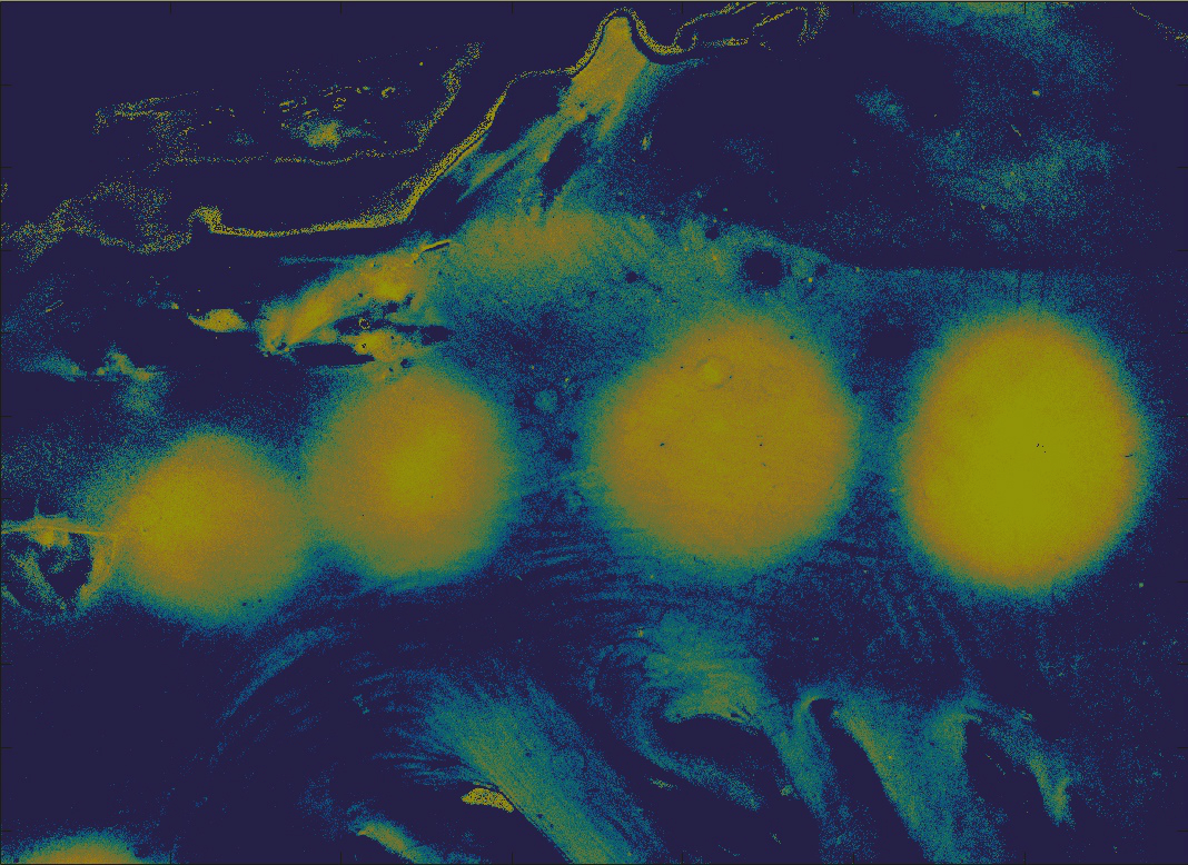
Segmentation results highlighted in blue of one patient using the automated segmentation algorithm currently in development.
The second group is focused on automatic breast segmentation to reduce the region we are looking at while searching for breast cancer tumors, by isolating the breasts from the rest of the image. In our algorithm, a canny edge detection technique is initially used to detect breast boundaries, which detects both weak and strong edges. An ellipse detection code searches the image for ellipses, which were most helpful for finding the inner curvature of the breasts. For most cases, we found the warmest regions of the images to be right below the lower curve of the breasts, which we used as another method to indicate where the breast edges are.
Using a point system, the different edges are weighted to determine the best fit to contain the desired pixels. The system accommodates various cases of breast sizes, since the effectiveness of different edge techniques used depended on breast size. Finally, the Laplacian of Gaussian edge detection technique is utilized to better display the edges outside of the body, as well as prominent breast boundaries. These are all compiled together, and the largest connected component is found (See image).
Current work focuses on detecting the curvature of the lines found, as well as enhancing the system to be completely automated in determining the most effective techniques for a given size and location of the breasts.