This past summer, many of our students had the opportunity to get their foot in the door of prestigious organizations, such as Singh Center for Nanotechnology, U.S Food and Drug Administration (FDA), and GW’s Nanotechnology Fellows Program. Here is what Caitlin, Nada and Zainab share about their accomplishments:
Caitlin Carfano:
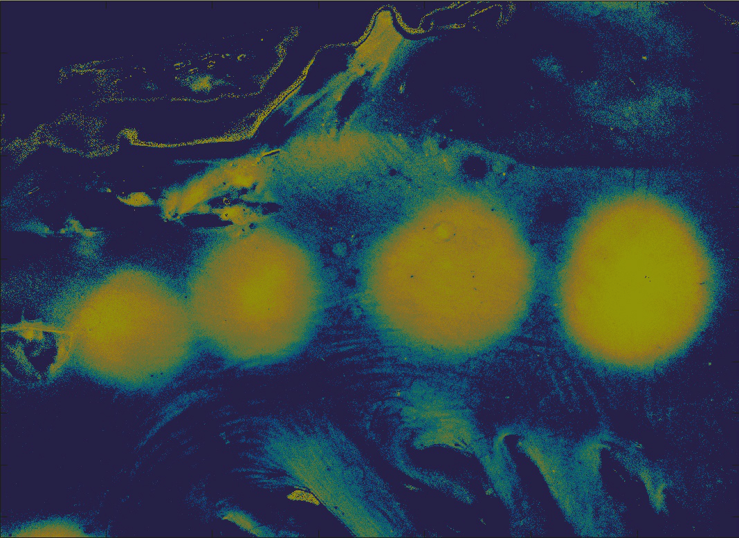
This summer I learned more than just information about the project I worked on, but also how to operate advanced equipment such as an Atomic Force Microscope. I regularly conducted research in the Singh Center for Nanotechnology surrounded by state of the art equipment and working with graduate students in my lab. I really enjoyed being able to work so closely with my mentor, Annemarie Exarhos, because I got to ask ample questions about research and I received numerous tips on creating and executing presentations. I also participated in a poster session symposium with other REU students and Penn scholars!
After learning about quantum technologies and how rapidly this field of study is advancing, I want to continue working in this field. Participating in this program also opened my eyes to all the other scientists and engineers doing quantum engineering research (I had a stimulating conversation with my eye doctor about quantum optics patents that he is working on!). I have always wanted to help others by pursuing a health/medical related career. After discovering this growing field and its incredible potential, I would be interested in research that applies quantum technologies to medical imaging devices.
Nada Kamona:
This summer, I was an ORISE Research Fellow at the U.S Food and Drug Administration (FDA) in the Division of Imaging, Diagnostics, and Software Reliability (DIDSR). Image analysis techniques, such as computer-aided diagnosis or radiomics, often rely on quantitative measurements of textures as input to characterize disease status. Ideally, texture features should discriminate different textures, and should be robust across a range of patient characteristics and image acquisition conditions. This study aims to identify such texture features through simulation. In my project, I worked on the variability of image texture quantification in simulated medical imaging systems, by examining the repeatability and reproducibility of 35 texture features to noise and spatial resolution using a library of textures that we generated. Not only has my positions allowed me to learn about quantitative imaging on a more in-depth and practical level but it has also taught me valuable critical thinking and problem solving skills. Working together with my supervisors at the FDA has helped me become a well-rounded scientist who is capable of both working by another’s direction and being an independent thinker.
Zainab Mahmood:
This past summer I conducted research under the Nanotechnology Fellows Program, a program designed to introduce students to nanotechnology, cutting-edge research, and GW’s new nano facilities. Through this program students attended lectures, seminars, and received hands-on cleanroom training. They received training on how to operate tools needed in nanofabrication and characterization processes such as the electron-beam lithography (EBL) tool, SEM, AFM, confocal microscope, probe station, and thermal evaporator. For my research project, I fabricated micro-scale gold contact design on graphene ribbons using Raith CAD software, electron beam lithography, oxygen plasma etching, and physical vapor deposition using a thermal evaporator and characterized electrical properties of graphene using four-point probe analysis and Raman spectroscopy.